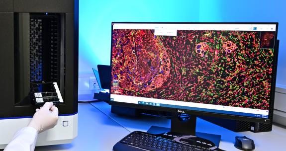
Things Are Getting Colorful: Tumor Research Using Multi-Stained Tissue Samples
A core task of pathology is the microscopic examination of surgical specimens after a histological slide has been prepared. The goal is to establish a diagnosis of the disease, which serves as the basis for treatment decisions. In cancer diagnostics, providing an individualized prognosis is often very difficult, as tumors represent a complex system of diseased and normal cells whose interactions are not yet fully understood.
As they grow, cancer cells interact with cells in the normal tumor microenvironment—particularly connective tissue cells and immune system cells. The type and function of the cells involved, as well as the extent of this interaction, determine the individual fate of the tumor and, ultimately, that of the patient.
Uncovering these cellular interactions is an essential step toward developing new diagnostic and therapeutic methods, as well as providing an individualized prognostic assessment. To achieve this, it is necessary to simultaneously visualize the different cell types in a histological specimen using specific stains. Multiplex immunofluorescence labeling (mIF) followed by multispectral imaging is an ideal method for this.
That is exactly what we will show you.
- We explain the complex cellular system in human carcinomas: tumor cells versus cells of the tumor microenvironment versus the extracellular tissue matrix
- We describe the preparation of conventional immunohistochemical sections for the detection of diagnostic marker proteins in tissue
- We describe the preparation of mIF sections and explain how they differ from conventional immunohistochemical sections used in routine pathological diagnostics.
- We demonstrate the microscopic visualization of mIF multiple labeling in carcinomas and explain the new insights that can be derived from the resulting “colorful images”
Further information is available on the Pathology Section’s website